Leg Bones Diagram ~ Tibia And Fibula Bone Anatomy. The muscles in the upper leg power many of our movements. The rounded, proximal end is the head of the femur, which articulates with the acetabulum of the hip bone to form the hip joint. (note, the radius and ulna bones also have this membrane.) this membrane keeps the tibia and fibula together and provides strength and stability for them. The bones of the leg and foot form part of the appendicular skeleton that supports the many muscles of the lower limbs. Most of the leg skeleton has bony prominences and margins that can be palpated and some serve as anatomical landmarks that define the extent of the leg.
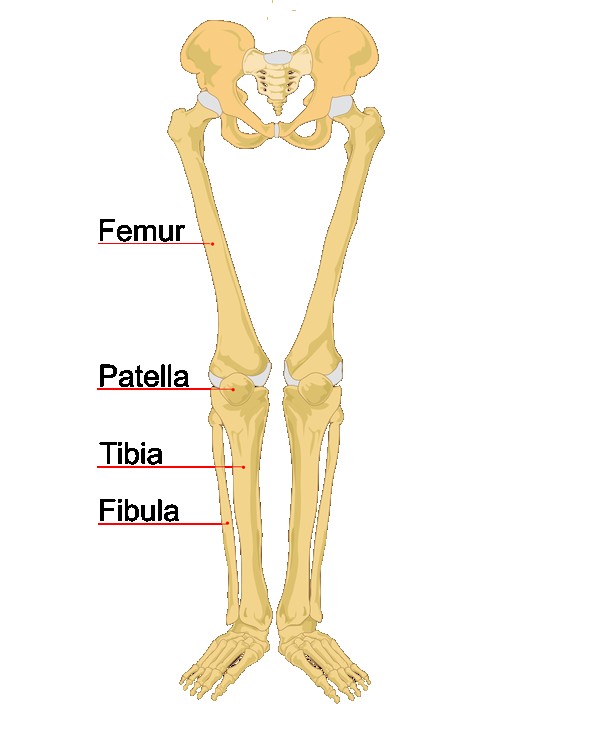
10 october 2007 (original upload date) source: Joints of hand anterior view, lateral view, right hand. The bones of the leg are the femur, tibia, fibula and patella.the foot bones shown in this diagram are the talus, navicular, cuneiform, cuboid, metatarsals and calcaneus. The lower leg extends from the knee to the ankle. The upper leg, in particular, is comprised of bones and muscles that are susceptible to injury, particularly when excess strain is placed upon them.
Diagram Upper Leg Bones Diagram Full Version Hd Quality Bones Diagram Diagramrt Ohimabrasserie It from bioluliaes.files.wordpress.com Now let's look at the tibia bone, which is the larger of the two leg bones, located medially. With different grades of sprains depending on severity. Then add shoulder blades, front legs. Joints of hand anterior view, lateral view, right hand. The patella is the kneecap bone. The bones together make up the hip. All four quadriceps muscles insert into the tibia (shin bone). The bones of the hip include the femur, the ilium, the ischium, and the pubis.
The patella is the kneecap bone.
The femur, or thigh bone, is the single bone of the thigh region (figure 6.51). Related posts of muscles and tendons of the leg muscle anatomy labeling. The patella is the kneecap bone. These muscles work together to produce movements such as standing, walking, running, and jumping. The lower leg is comprised of two bones, the tibia and the smaller fibula. It lies within the quadriceps tendon. 10 october 2007 (original upload date) source: The rounded, proximal end is the head of the femur, which articulates with the acetabulum of the hip bone to form the hip joint. Last add pelvis, back leg (part of one bag leg missing). These bones have a marrow, but not a bone marrow cavity. Lower jaw (mandible) collar bone. This large tendon from the powerful thigh muscles (quadriceps) wraps round the patella and is attached to the top of the lower leg bone (tibia). Disposition of rotator cuff muscles diagram.
The femur, or thigh bone, is the single bone of the thigh region (figure 6.51). Your leg bones are very large and strong to help support the weight of your body. This image is an edited version of this image that was created by user:ladyofhats (mariana ruiz villarreal). Diagram and names of leg bones, diagram of foot and leg bones, diagram of leg bones, diagram of lower leg bones, diagram of the bones in your leg, bone, diagram and. The bones of the hip include the femur, the ilium, the ischium, and the pubis.
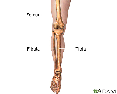
Leg Skeletal Anatomy Medlineplus Medical Encyclopedia Image from medlineplus.gov All four quadriceps muscles insert into the tibia (shin bone). Then add shoulder blades, front legs. The thigh bone, or femur, is the large upper leg bone that connects the lower leg bones (knee joint) to the pelvic bone (hip joint). Framework of bones, class 6. These muscles work together to produce movements such as standing, walking, running, and jumping. Diagram and names of leg bones, diagram of foot and leg bones, diagram of leg bones, diagram of lower leg bones, diagram of the bones in your leg, bone, diagram and. The tibia and fibula are two long bones that run parallel to each other, forming the scaffold of the leg and providing attachment points for many muscles. The tibia and the fibula, at the top of the ankle joint.
Likely shrew, mouse, vole or rat.
The upper leg, in particular, is comprised of bones and muscles that are susceptible to injury, particularly when excess strain is placed upon them. Related posts of muscles and tendons of the leg muscle anatomy labeling. Framework of bones, class 6. The thigh bone, or femur, is the large upper leg bone that connects the lower leg bones (knee joint) to the pelvic bone (hip joint). Likely shrew, mouse, vole or rat. The diagram of bones in the ankle and foot is given below: The bones together make up the hip. Now let's look at the tibia bone, which is the larger of the two leg bones, located medially. The bones of the leg are the femur, tibia, fibula and patella.the foot bones shown in this diagram are the talus, navicular, cuneiform, cuboid, metatarsals and calcaneus. In this small section, we'll briefly mention the main parts of the leg, namely the bones, muscles, and neurovasculature. The quadriceps muscle attachment points. Then add shoulder blades, front legs. These muscles work together to produce movements such as standing, walking, running, and jumping.
Related posts of muscles and tendons of the leg muscle anatomy labeling. This image is an edited version of this image that was created by user:ladyofhats (mariana ruiz villarreal). Now let's look at the tibia bone, which is the larger of the two leg bones, located medially. It also separates muscles on the anterior and posterior parts of the leg. This large tendon from the powerful thigh muscles (quadriceps) wraps round the patella and is attached to the top of the lower leg bone (tibia).
Leg Definition Bones Muscles Facts Britannica from cdn.britannica.com The upper leg, in particular, is comprised of bones and muscles that are susceptible to injury, particularly when excess strain is placed upon them. The bones of the leg and foot form part of the appendicular skeleton that supports the many muscles of the lower limbs. The two main bones of the leg are the tibia ('shin bone') located medially and the fibula, which is located more laterally. The tibia, commonly known as the 'shin bone', is the largest and most medial of the two.you can palpate its anterior border when you run your finger down the anterior aspect of your leg. Also called the shin bone, the tibia is the longer of the two bones in the. Related posts of muscles and tendons of the leg muscle anatomy labeling. The bones of the hip include the femur, the ilium, the ischium, and the pubis. At the same time, the bones and joints of the leg and foot must be strong enough to support the body's weight while remaining.
Labeled human leg bones created for use in leg bone.
The quadriceps muscles straighten the knee. Disposition of rotator cuff muscles diagram. There are in all 7 bones, which fall under tarsal bones category. With different grades of sprains depending on severity. The bones together make up the hip. Then add shoulder blades, front legs. Most of the leg skeleton has bony prominences and margins that can be palpated and some serve as anatomical landmarks that define the extent of the leg. Discuss who might have taken the leg away (bobcat, coyote, fox, bald eagle) and imagine the scenario of a scavenger finding this big meal! The tibia is the largest of the two, hence it is responsible for weight bearing. The lower leg is comprised of two bones, the tibia and the smaller fibula. Related posts of muscles and tendons of the leg muscle anatomy labeling. The femur, or thigh bone, is the single bone of the thigh region (figure 6.51). Muscle anatomy labeling 12 photos of the muscle anatomy labeling anatomy muscle labeling games, holes anatomy muscle labeling, mcgraw hill anatomy muscle labeling, muscle anatomy model labeled, skeletal muscle anatomy labeling, human muscles, anatomy muscle labeling games, holes anatomy muscle labeling, mcgraw hill.